Far-infrared Therapy as a Novel Treatment for Encapsulating Peritoneal Sclerosis
Letters to the Editor
......A 45-year-old man with a medical history of end-stage renal disease was admitted owing to abdominal fullness, vomiting, and poor appetite for 1 month. The patient had received 13 years of PD followed by removal of the PD catheter owing to refractory peritonitis, resulting in a shift to hemodialysis 2 months before this admission. A physical examination showed a body temperature of 36.5 °C, a
distended abdomen and hypoactive bowel sounds. Th e dialysate effluent was dark red in appearance. Laboratory examinations
revealed normal blood leukocyte count (WBC: 7,100/mm3 ) but an elevated C-reactive protein level (50.3 mg/l). An abdominal radiograph was obtained to evaluate his symptoms. The intestinal wall was outlined by continuous calcifications (Figure 1 , arrows), and the curvilinear calcification on the right side of the radiograph was peritoneal calcification (Figure 1, white arrows). A contrast enhanced
computed tomography scan demonstrated fluid within the abdomen surrounded by thickened, enhancing peritoneum. The adherent bowel loops were collected centrally by the encapsulating calcified peritoneum, supporting the diagnosis of EPS (Figure 2, white
arrows).
Aft er 3 months of far-infrared therapy, his bowel movement improved. He was able to tolerate a regular oral diet and the color of the dialysate effluent became lightly pink. Because of the improvement in the symptoms aft er far-infrared therapy, our patient decided to follow a conservative treatment plan. Follow-up contrast-enhanced computed tomography scan showed an improvement in dilated loops of small bowel (Figure 3, white arrows). Remarkably, his nutritional status also improved with a significant increase of serum albumin level from 2.7 to 4.2 g/dl, as well as body weight from 59.1 to 62.9 kg aft er far-infrared therapy for 12 months....
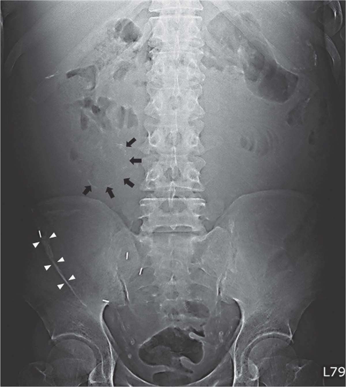
Figure 1.
An abdominal radiograph showed
intestinal wall was outlined by continuous calcifications (arrows), and the curvilinear calcifi cation on the right side of the radiograph was peritoneal calcifi cation (white arrows).
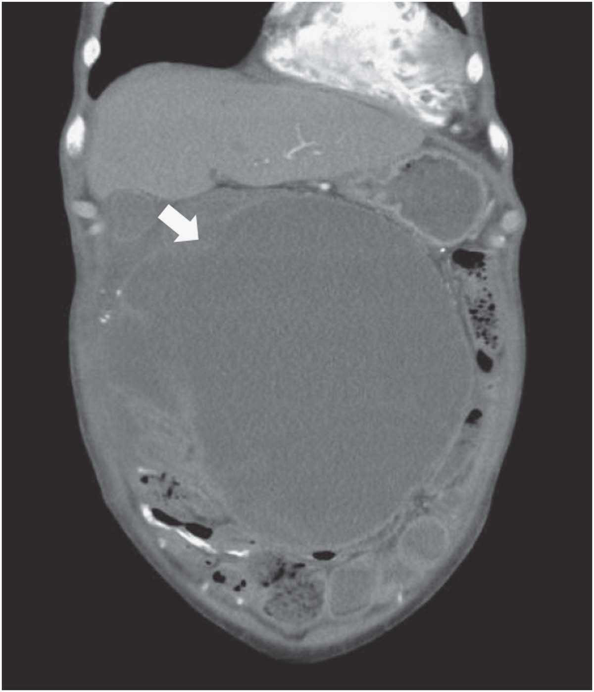
Figure 2.
A contrast-enhanced computed tomography scan demonstrated that the adherent bowel loops were collected centrally by the encapsulating calcifi ed peritoneum, supporting the diagnosis of encapsulating peritoneal sclerosis (EPS) (white arrows).
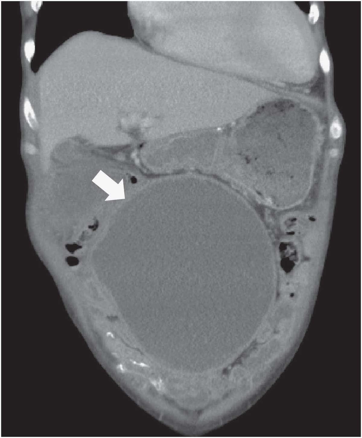
Figure 3.
Follow-up computed tomography scan
showed an improvement in dilated loops of small bowel (white arrows).
-Am J Gastroenterol. 2014 Dec;109(12):1957-9.
Want to know more?
Contact us for more detail